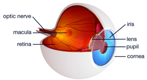
About the Eye


The eye itself is approximately the size of a ping pong ball, about one and a quarter inches in diameter. If we were to cut an eye in half, the back half is lined with a very thin tissue. This tissue is the retina of the eye. The retina is about 1/10th of the thickness of a piece of saran wrap, has 10 layers, and contains 125 million cells. When light strikes a retinal receptor cell it creates an electrical impulse that travels through the appropriate layers of the retina and then along a fiber of the optic nerve back to the brain. The brain may receive hundreds to tens of thousands of messages and by integrating them together it creates images, which is what we know as sight. Therefore, sight takes place in the brain, not in the eye.
In the center of the retina there is a small area about one and a half millimeters in diameter, about the size of the head of a pin. This area contains the greatest concentration of cells in the retina and is know as the Macula.
The macula serves three major functions:
- Direction – When we look directly at an object the eye and brain coordinate together and the brain aims the macula directly at what we are viewing. The object we are looking at represents “central vision”. Once the brain knows what is in the center it knows what surrounds it as well, which we call “peripheral vision”.
- Fine Detail – The macula has a high concentration of cells and is the most sensitive part of the retina relative to detail and as such it is responsible for functions like reading, writing, playing cards, driving, and identifying faces.
- Color – Most of the “cone” cells are in the macula as well. These are the cells that are responsible for most of our ability to see and identify color.
When we think of the retina projected out into the world we think of it as being a “bulls-eye” target, where the bulls-eye is the macula, and each ring away from the macula we go on the target represents a zone with fewer cells. When we reach the final ring we are at the equator of the retina which is blind, having no receptive cells, and this is normal. From there the closer we return to the center, the more cells we have, and the more sensitive the retina becomes, until we return to the macula, which again, is the most sensitive of all.


